alpha-1-antichymotrypsin: interplay with chymotrypsin-like proteinases
Abstract
The interplay of human plasma alpha-1-antichymotrypsin with serine proteinases from totally different tissues has been investigated. The protein was discovered to kind steady complexes with pancreatic chymotrypsin, leukocyte cathepsin G, and mast cell chymotrypsin. No inhibition of pancreatic trypsin or leukocyte elastase may very well be demonstrated. With mixtures containing each alpha-1-antichymotrypsin and alpha-1-proteinase inhibitor, it was discovered that the previous preferentially inactivated leukocyte cathepsin G, whereas the latter confirmed a robust choice for pancreatic chymotrypsin.
However, leukocyte elastase was particularly inactivated by alpha-1-proteinase inhibitor even in 1:1 mixtures with chymotrypsin. All of those outcomes taken collectively recommend that one of many main capabilities of alpha-1-antichymotrypsin is to inactivate leukocyte cathepsin G, whereas alpha-1-proteinase inhibitor controls the exercise of different serine proteinases, significantly leukocyte elastase.
CYTOMORPHOLOGY OF SOLID-PSEUDOPAPILLARY NEOPLASM:
- • extremely mobile aspirate
- • myxoid or hyalinized vascular stalks lined by neoplastic cells
- • delicate granular cytoplasm, vague cell borders
- • spherical or oval nuclei
- • nuclear grooves
- • inconspicuous nucleoli
- • foam cells, necrotic particles
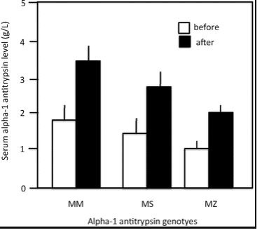
antitrypsin genotypes
FNA yields extremely mobile smears composed of a monotonous inhabitants of cuboidal cells organized in loosely cohesive teams, as remoted cells, and, most characteristically, as a single or a number of layer round vascular constructions which can be typically thickened by a myxoid or hyaline materials (Fig. 13-7A). The tumor cells have delicate granular cytoplasm with vague cell borders. The nuclei are spherical to oval with finely dispersed chromatin, clean or grooved nuclear contours, and vague nucleoli (Fig. 13-7B). Mitotic figures are often inconspicuous. The background might include considerable blood, foam cells, globules of amorphous myxoid materials, and necrotic particles.
[Linking template=”default” type=”products” search=”antitrypsin genotypes” header=”1″ limit=”23″ start=”3″ showCatalogNumber=”true” showSize=”true” showSupplier=”true” showPrice=”true” showDescription=”true” showAdditionalInformation=”true” showImage=”true” showSchemaMarkup=”true” imageWidth=”” imageHeight=””]