DESCRIPTIONApplication
Packaging
Biochem/physiol Actions
Preparation Note
Analysis Note
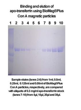
BioMag Concanavalin A
PRODUCT DESCRIPTION
Concanavalin A (Con A) is a extensively used lectin that selectively binds to the glycoproteins, a-mannopyranosyl and a-glucopyranosy. These are generally discovered within the cell wall of yeast and fungi, and the cell membrane of mammalian cells and tissues.
- Stain the cell wall of yeast and fungi, and the cell membrane of mammalian cells and tissues
- Detect glycoconjugates in microscopy and move cytometry
- Stain glycoproteins in gels
- Withstands fixation and permeabilization
- Choice of 10 CF dyes from UV to near-infrared
- Superior CF dyes are brilliant, photostable, and water-soluble
Lectins are additionally versatile probes for detecting glycoconjugates in microscopy and move cytometric purposes and for gel staining of glycoproteins. In impartial and alkaline options, Con A exists as a tetramer with a molecular weight of roughly 104 kDa. In acidic options (pH under 5.0), Con A exists as a dimer. Con A can be utilized to selectively stain the cell floor of dwell cells, and face up to fixation and permeabilization. When cells are mounted and permeabilized earlier than staining, fluorescent lectins stain each cell floor and organelles within the secretory pathway.
Find the Right Stain for Your Application
Con A and different lectins are carbohydrate binding proteins that acknowledge particular sugar moieties on glycoproteins. The presence and distribution of those targets range between cell varieties and tissues. As a consequence, different cell floor stains or different lectin conjugates, Wheat Germ Agglutinin (WGA) Conjugates and PNA Lectin Conjugates, could produce higher floor staining and could also be extra applicable to your cell kind. Lectin conjugates can be utilized to selectively stain the cell floor of dwell cells, and face up to fixation and permeabilization. When cells are mounted and permeabilized earlier than staining, fluorescent lectins stain each cell floor and organelles within the secretory pathway.
[Linking template=”default” type=”products” search=”Concanavalin A” header=”1″ limit=”26″ start=”1″ showCatalogNumber=”true” showSize=”true” showSupplier=”true” showPrice=”true” showDescription=”true” showAdditionalInformation=”true” showImage=”true” showSchemaMarkup=”true” imageWidth=”” imageHeight=””]